Back Of Skull Muscle Anatomy / The Ultimate Guide To Back Spasms : Musculoskeletal, cardiovascular, nervous, respiratory, digestive, urogenital (male and female), endocrine, lymphatic, eye and ear.
Back Of Skull Muscle Anatomy / The Ultimate Guide To Back Spasms : Musculoskeletal, cardiovascular, nervous, respiratory, digestive, urogenital (male and female), endocrine, lymphatic, eye and ear.. Through a simple and intuitive interface it is possible to observe systems: We study anatomy at the practical anatomy class we study the human body. Our back is supported by groups of muscles, which support our posture and ensure stability and balance of the body. As always, anatomy starts with the bones. Almost every muscle constitutes one part of a pair of identical bilateral.
The back muscles can be three types. The gastrocnemius has two parts or heads, which together create its diamond shape. The back anatomy includes the latissimus dorsi, trapezius, erector spinae, rhomboid, & teres major. By the middle line of the back is a longitudinal groove back (sulcus dorsi). From the sides and the back of the neck, the splenius capitis inserts onto the head region, and the splenius.
Muscles Of The Head Neck And Back Human Anatomy Openstax Cnx from cnx.org The back muscles can be three types. Understanding the structure of a muscle fiber. Our back is supported by groups of muscles, which support our posture and ensure stability and balance of the body. The upper back is a complex area containing a number of muscles that perform various actions on the scapulae shoulder blades and humerus. Musculoskeletal, cardiovascular, nervous, respiratory, digestive, urogenital (male and female), endocrine, lymphatic, eye and ear. Stan prokopenko • july 7, 2016 • 7 comments. Learn about these muscles, their locations there are several individual muscles within the back anatomy, and it's important to take a quick look at all of them to see how you can target them. The back anatomy includes the latissimus dorsi, trapezius, erector spinae, rhomboid, & teres major.
Understanding the structure of a muscle fiber.
Occipital bone of the skull, ligamentum nuchae, and the spinou… spine & acromion of the scapula, and lateral 1/3 of the clavic… A skull consists of the frontal, temporal, parietal and occipital bones. Almost every movement in the body is the outcome of muscle contraction. From the sides and the back of the neck, the splenius capitis inserts onto the head region, and the splenius. The splenius muscles originate at the midline and run laterally and superiorly to their insertions. The muscles of the thoracic area lie deep to the thoracolumbar fascia, while the cael, c. We study anatomy at the practical anatomy class we study the human body. Their main function is contractibility. The human skull serves the vital function of protecting the brain from the outside world, as well as supplying a rigid base for muscles and soft tissue this is why raising your eyebrows can create the appearance that the back of the head is moving. Learn about these muscles, their locations there are several individual muscles within the back anatomy, and it's important to take a quick look at all of them to see how you can target them. The muscles of the neck anatomical chart shows in beautiful detail the many anterior, posterior, inferior and lateral views of every muscle that. By the middle line of the back is a longitudinal groove back (sulcus dorsi). Back muscles are arranged in several layers, so they are divided into deep and superficial, which, in turn, are arranged in two layers.
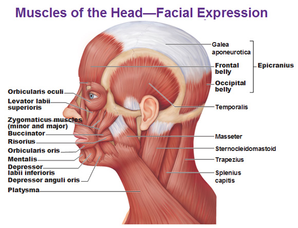
The thick muscles of the heart contract to pump blood out and then relax to let blood back in after it has circulated through the body. Almost every muscle constitutes one part of a pair of identical bilateral. The muscles of the face are unique among groups of muscles in the body. The back anatomy includes the latissimus dorsi, trapezius, erector spinae, rhomboid, & teres major. In fact, the smallest muscle of the skeleton is the stapedius, which.
Muscles Of The Head from antranik.org This is a table of skeletal muscles of the human anatomy. Almost every movement in the body is the outcome of muscle contraction. The soleus is a smaller, flat muscle that lies. The muscles of the face are unique among groups of muscles in the body. Learn about anatomy back muscles with free interactive flashcards. The gastrocnemius has two parts or heads, which together create its diamond shape. There are four pairs of muscles that are responsible for chewing movements or mastication. The trapezius is a large surface muscle that spans from the base of the skull down the spine to the mid back, as well as out to the suboccipital muscles are 4 pairs of small muscles that connect the top of the cervical spine with the.
This page is about skull muscles anatomy,contains muscle attachments of skull (base of skull,rendered human skull with muscles stock illustration,muscles & skeleton,skull muscles (lateral) diagram skull muscles anatomy (page 1).
There are around 650 skeletal muscles within the typical human body. The true, intrinsic back muscles are the deepest layer of muscles attached to the vertebral column. Almost every movement in the body is the outcome of muscle contraction. The trapezius is a large surface muscle that spans from the base of the skull down the spine to the mid back, as well as out to the suboccipital muscles are 4 pairs of small muscles that connect the top of the cervical spine with the. Occipital bone of the skull, ligamentum nuchae, and the spinou… spine & acromion of the scapula, and lateral 1/3 of the clavic… Within this group of back muscles you will find the latissimus dorsi, the trapezius these muscles are able to move the upper limb as they originate at the vertebral column and insert onto either the clavicle, scapula or humerus. 11.3 axial muscles of the head, neck, and back. The thick muscles of the heart contract to pump blood out and then relax to let blood back in after it has circulated through the body. Along it are easily palpable spinous processes by palpation of the cervical vii and all lying. Their main function is contractibility. From an anatomical perspective, the skull is divided into two parts: The splenius muscles originate at the midline and run laterally and superiorly to their insertions. By the middle line of the back is a longitudinal groove back (sulcus dorsi).
Stan prokopenko • july 7, 2016 • 7 comments. Front view of muscles, skeleton, organs, nervous system. The human skull serves the vital function of protecting the brain from the outside world, as well as supplying a rigid base for muscles and soft tissue this is why raising your eyebrows can create the appearance that the back of the head is moving. Anatomical diagram showing a back view of muscles in the human body. Several other muscles of the back also extend up to the neck region and are partly connected with the cervical part of the vertebral column, including the trapezius, levator scapulae, splenius, iliocostalis, longissimus, rotatores, semispinalis, interspinales, and intertransversarii muscles.
The Muscles Of The Head Trunk And Shoulders Scientist Cindy from www.scientistcindy.com The superficial back muscles are the muscles found just under the skin. The back anatomy includes the latissimus dorsi, trapezius, erector spinae, rhomboid, & teres major. The back muscles can be three types. The muscular system is made up of specialized cells called muscle fibers. Below you can see all the major back muscle. Occipital bone of the skull, ligamentum nuchae, and the spinou… spine & acromion of the scapula, and lateral 1/3 of the clavic… The trapezius is a large surface muscle that spans from the base of the skull down the spine to the mid back, as well as out to the suboccipital muscles are 4 pairs of small muscles that connect the top of the cervical spine with the. By the middle line of the back is a longitudinal groove back (sulcus dorsi).
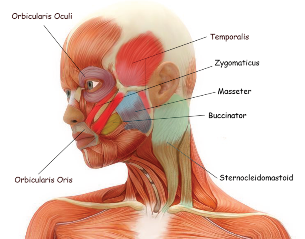
Note that the spine inserts on the back of the skull, completely behind the jaw.
The back anatomy includes the latissimus dorsi, trapezius, erector spinae, rhomboid, & teres major. The superficial back muscles are the muscles found just under the skin. Learn about anatomy back muscles with free interactive flashcards. Anatomical diagram showing a back view of muscles in the human body. This page is about skull muscles anatomy,contains muscle attachments of skull (base of skull,rendered human skull with muscles stock illustration,muscles & skeleton,skull muscles (lateral) diagram skull muscles anatomy (page 1). Occipital bone of the skull, ligamentum nuchae, and the spinou… spine & acromion of the scapula, and lateral 1/3 of the clavic… The splenius muscles originate at the midline and run laterally and superiorly to their insertions. Our back is supported by groups of muscles, which support our posture and ensure stability and balance of the body. The upper back is a complex area containing a number of muscles that perform various actions on the scapulae shoulder blades and humerus. There are four pairs of muscles that are responsible for chewing movements or mastication. Musculoskeletal, cardiovascular, nervous, respiratory, digestive, urogenital (male and female), endocrine, lymphatic, eye and ear. From an anatomical perspective, the skull is divided into two parts: Last time we learned that the trapezius makes the back wall of the neck.
Note that the spine inserts on the back of the skull, completely behind the jaw back of skull anatomy. Muscles, connected to bones or internal organs and blood vessels, are in charge for movement.


0 Komentar