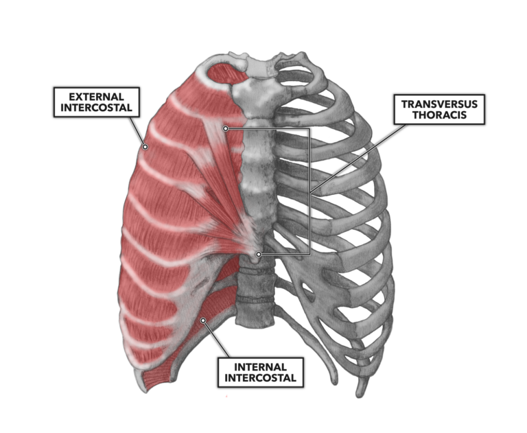
Anatomy Of Rib Cage Muscles : Thoracic Spine / For more anatomy content please follow us and visit our website:. Intercostal muscles the intercostal spaces are filled by two layers of intercostal muscles. Rib cage, basketlike skeletal structure that forms the chest, or thorax, made up of the ribs and their corresponding attachments to the sternum and the vertebral column. This is a stereogram, to be viewed in crossview technique. The ribs are curved, flat bones which form the majority of the thoracic cage. Check out our muscle anatomy reference charts to learn faster!
The major abdominal muscles include the transverse according to medical news today, it's important to understand the anatomy of the rib cage when determining whether pain under the rib cage is mild. In the back, latissimus dorsi and erector spinae muscles (anatomy lesson #10) cover the 11th and 12th ribs of the thoracic cage and deeper yet are the paired abdominal kidneys flanking the. Contributing to their role in protecting the internal thoracic organs. The thoracic cage is part of the axial skeleton (also known as the rib cage), and consists of 24 ribs, the sternum, costal cartilage, and the 12 thoracic vertebrae. Animal physiotherapy foundation programme , the first of two courses on the equine forelimb.
The Intercostal Muscles of the Ribcage from corewalking.com Contributing to their role in protecting the internal thoracic organs. Check out our muscle anatomy reference charts to learn faster! In the back, latissimus dorsi and erector spinae muscles (anatomy lesson #10) cover the 11th and 12th ribs of the thoracic cage and deeper yet are the paired abdominal kidneys flanking the. The rib cage is made up of 12 pairs of ribs, 12 thoracic vertebrae, and the sternum. Abdomen & ribs muscle movements. Rendering done with a carestream workstation. Everyone has nice muscles in ct scanning! Animal physiotherapy foundation programme , the first of two courses on the equine forelimb.
Muscles that move the rib cage attach to the rib cage.
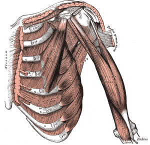
The rib cage is the arrangement of ribs attached to the vertebral column and sternum in the thorax of most vertebrates, that encloses and protects the vital organs such as the heart, lungs and great vessels. Rendering done with a carestream workstation. Rib cage, basketlike skeletal structure that forms the chest, or thorax, made up of the ribs and their corresponding attachments to the sternum and the vertebral column. All muscles that are attached to the human rib cage have the. The ribs are curved, flat bones which form the majority of the thoracic cage. The intercostal muscles extend from the vertebrae behind to the sternum in front. Seventeen muscles attach to the scapula, and it articulates with the clavicle to form the shoulder girdle or pectoral girdle, which. Anatomy the rib cage is a bony structure found in the chest thoracic cavity. Muscles that move the rib cage attach to the rib cage. During normal breathing, contraction of the major inspiratory muscle, the diaphragm, produces both rib cage expansion and a downward movement of the diaphragm. Another important feature of the rib cage is the manubriosternal joint also known as the sternal angle of louis. Everyone has nice muscles in ct scanning! The thoracic cage (rib cage) is the skeletal framework of the thoracic wall, which encloses the thoracic cavity.
The following general rules regarding actions can be. Muscles of thorax, upper extremities, back and diaphragm are given connection by this twelve pairs of ribs are attached to the thoracic vertebrae. The rib cage surrounds the lungs and the heart, serving as an important means of bony protection for these vital organs. The other attachment of these muscles is usually considered to be either superior or inferior to the rib attachment. For more anatomy content please follow us and visit our website:
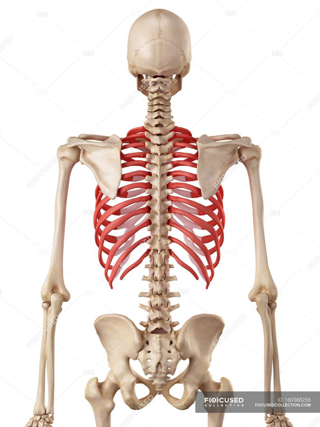
Human rib cage anatomy — human physiology, osteology ... from st.focusedcollection.com See more ideas about anatomy, rib cage anatomy, anatomy study. Ribs & thoracic cage muscles attachments. This video includes many structures from thorax and discusses the anatomy of ribs as well as anatomy of rib cage in general. The fibers attach to the rib cage and the pubis of the hip bones. During normal breathing, contraction of the major inspiratory muscle, the diaphragm, produces both rib cage expansion and a downward movement of the diaphragm. The thoracic cage consists of the 12 thoracic vertebrae, the associated intervertebral discs, 12 pairs of ribs with their costal cartilages, and the sternum. 'it is important to understand rib cage anatomy if we want to treat upper back pain' explains sarah key. Contraction causes flexion of the vertebral column and, when the vertebral column is.
Some of the most common causes of rib cage pain stem from sports and physical activity.
The thoracic cage (rib cage) is the skeletal framework of the thoracic wall, which encloses the thoracic cavity. Struggling with learning muscle attachments? These muscles may be located anteriorly, posteriorly, and/or laterally. The fibers attach to the rib cage and the pubis of the hip bones. The thoracic cage is part of the axial skeleton (also known as the rib cage), and consists of 24 ribs, the sternum, costal cartilage, and the 12 thoracic vertebrae. All muscles that are attached to the human rib cage have the. Check out our muscle anatomy reference charts to learn faster! See more ideas about anatomy, rib cage anatomy, anatomy study. The rib cage surrounds the lungs and the heart, serving as an important means of bony protection for these vital organs. 'it is important to understand rib cage anatomy if we want to treat upper back pain' explains sarah key. This is a stereogram, to be viewed in crossview technique. Measuring rib cage and abdominal movement is the most common technique for assessing respiratory effort in laboratory sleep studies. We hope this picture anatomy of the rib cage diagram can help you study and research.
The other attachment of these muscles is usually considered to be either superior or inferior to the rib attachment. Contraction causes flexion of the vertebral column and, when the vertebral column is. The rib cage surrounds the lungs and the heart, serving as an important means of bony protection for these vital organs. Originate at the lower border of the rib inserting into the superior border of the rib below. Muscles that move the rib cage attach to the rib cage.
Anatomy Of Rib Muscles - Anatomy Drawing Diagram from www.crossfit.com The thoracic spine supports twelve pairs of ribs that slope gently down from the back as they pass around to encase the thorax. Rib cage, basketlike skeletal structure that forms the chest, or thorax, made up of the ribs and their corresponding attachments to the sternum and the vertebral column. Originate at the lower border of the rib inserting into the superior border of the rib below. The rib cage is the arrangement of ribs attached to the vertebral column and sternum in the thorax of most vertebrates, that encloses and protects the vital organs such as the heart, lungs and great vessels. The primary responsibilities of the ribcage involve protecting the thoracic visceral organs, enclosing the thoracic visceral organs, and is included in the general mechanics of the process of breathing. Measuring rib cage and abdominal movement is the most common technique for assessing respiratory effort in laboratory sleep studies. But for an anatomy study its not. Muscle spasms felt within the rib cage may also be caused by the abdominal muscles.
Abdomen & ribs muscle movements.
The thoracic cage (rib cage) is the skeletal framework of the thoracic wall, which encloses the thoracic cavity. Muscles that move the rib cage attach to the rib cage. These muscles may be located anteriorly, posteriorly, and/or laterally. Abdomen & ribs muscle movements. Contributing to their role in protecting the internal thoracic organs. Seventeen muscles attach to the scapula, and it articulates with the clavicle to form the shoulder girdle or pectoral girdle, which. Struggling with learning muscle attachments? Muscles are often named for their primary action. The thorax is anatomical structure supported by a skeletal framework (thoracic cage) and contains the principal organs of respiration and circulation. The primary responsibilities of the ribcage involve protecting the thoracic visceral organs, enclosing the thoracic visceral organs, and is included in the general mechanics of the process of breathing. Muscle spasms felt within the rib cage may also be caused by the abdominal muscles. Animal physiotherapy foundation programme , the first of two courses on the equine forelimb. This is a stereogram, to be viewed in crossview technique.
Muscles are often named for their primary action anatomy of rib cage. Muscle spasms felt within the rib cage may also be caused by the abdominal muscles.


0 Komentar